T-Wave Alternans To evaluate patients for risk of the serious arrhythmia that causes sudden cardiac death, this treadmill stress test is performed. This test records microscopic changes in the T-Wave portion your ECG. Changes in the T wave has been shown to be effective in determining patients who are who are at high risk for sudden death.
________________________________________
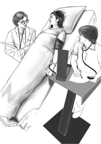
Tilt table study To evaluate patients with unexplained dizziness and fainting spells (syncope), this simple and noninvasive test is performed. Specific diagnoses such as carotid hypersensitivity syndrome ,vasovagal syncope, postural orthostatic tachycardia syndrome, and neurocardiogenic syncope can be made from this study. After an overnight fast, the test is usually done on an outpatient setting. The patient is strapped to a tilt table to prevent accidentally falling and tilted to a 75 degree angle and monitored with ECG and blood pressure recording continuously for 20 to 45 minutes. The underlying cause of fainting can be revealed in about 50-75% of the cases for patients with recurring fainting spells. To confirm the diagnosis, the fainting or near-fainting episodes must be reproduced during the test.
________________________________________
Electrophysiology study (EP Study) To evaluate patients whose cause for fainting or severe palpitations remains unknown despite extensive noninvasive evaluations, this invasive study is generally performed. It is also useful to differentiate the numerous causes for an arrhythmia, risk-stratify patients with known or suspected arrhythmias, and eliminate the culprit of the arrhythmia when used in conjunction with radiofrequency ablation catheters.
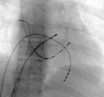
An EP study is performed by an electrophysiologist under fluoroscopy guidance in the cardiac catheterization laboratory, where coronary angiograms and angioplasties are performed. Several EP catheters are inserted through the femoral vein in the groin into the heart. Then electrical stimulation of the heart is performed through these EP catheters by the Electrophysiologist. Slow heartbeat or heart block and reproducing the cause of a rapid arrhythmia which the patient chronically suffers from, can reveal the underlying electrical conduction problem. Therefore, patients are not usually in their arrhythmia at the time of the procedure.
The electrophysiologist can prescribe a pacemaker if a slow heartbeat is documented. Depending on the type of rapid heartbeat discovered, different treatment options are possible. For life threatening, ventricular tachycardia an implantable defibrillator is indicated. For the more benign form of rapid heartbeats, such as supraventricular tachycardia, the arrhythmia can be “mapped” to determine their cause and then a cardiac ablation can be performed. The majority of the time, cardiac EP ablations can successfully eliminate the culprit of the arrhythmias, resulting in a long-term permanent cure. Thus, an Electrophysiology study is a diagnostic study that evaluates the electrical health of the heart as opposed to a coronary angiogram that is designed to look for clotted arteries of the heart (coronary arteries). Electrophysiologists can steer the EP platinum tip of electrophysiology catheters by turning the proximal handle. This allows Electrophysiologists to place the EP catheters in the perfect spot.
Many patients are very reluctant to have a test which can provoke their arrhythmias; especially, when the symptoms are serious symptoms like their rapid heartbeat or fainting. Unfortunately, the only way to confirm the causes of their arrhythmias in most patients may be reproducing the arrhythmia in an EP lab. Luckily, there is no safer place to have an arrhythmia than in the cardiac catheterization laboratory under the direct care of a Cardiac Electrophysiologist and in the presence of an entire team of personnel specializing in the chronic and emergency management of arrhythmias.
 to Diagnose Arrhythmias Part III of IV)